DNA Has Another Zipper, and It Joins Two Complete Double Helices
Atomic-force microscopy revealed two intact DNA double helices aligned over several turns. Simulations suggest positively charged ions help bridge them, but the experiment was a structural snapshot, not a movie of DNA pairing inside a cell.

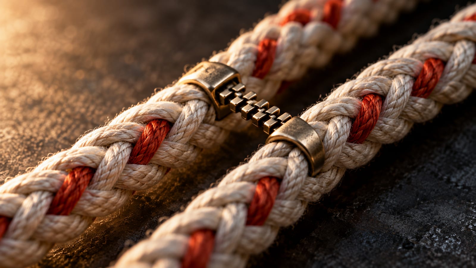
Forget, for a moment, the DNA zipper from biology class, the one in which two strands separate and matching bases reconnect.
This is a different zipper altogether.
Researchers have imaged two already-complete DNA double helices pressed alongside one another. In the sharpest images, their spiral grooves lined up over several turns, even though both molecules carried electrical charges that should have pushed them apart.
The structures offer striking support for an idea physicists have pursued for 25 years: positively charged ions can arrange themselves around DNA in ways that let one double helix interlock electrostatically with another.
There is an important qualification. The researchers did not film a zipper racing shut inside a living cell. They obtained topographic snapshots of short DNA molecules deposited on mica and submerged in liquid. The proposed molecular bridges came from atom-by-atom computer simulations, because the microscope could not resolve individual ions.
Even so, the combination exposes a strange possibility with unusual clarity. DNA may be capable of making organized contact with other DNA before a protein reads either molecule.
The problem with putting DNA next to DNA
The outside of a DNA double helix is lined with phosphate groups, which give it a negative charge. Put two such molecules near each other and ordinary electrostatics says they should repel, like matching poles of two magnets.
Cells nevertheless pack enormous lengths of DNA into compact spaces. Chromosomes must also find related regions during processes such as homologous recombination, when genetic information is exchanged or used for repair. Proteins perform much of that work, but experiments have repeatedly suggested that intact double helices can show a preference for homologous sequences without the usual recombination machinery conducting the initial comparison.
That leaves a basic question: How could two repelling molecules get close enough to recognize anything?
A possible answer arrived in 2001, when physicists Alexei Kornyshev and Sergey Leikin proposed that DNA’s charge is not distributed like a featureless negative tube. A helix has twisted backbones, major and minor grooves, and sequence-dependent variations in shape. Counterions gathering around it could therefore create a patterned electrical surface.
Two helices with compatible patterns might lower their energy by aligning those patterns, a little like meshing two spiral staircases. Sequence-dependent distortions could then help homologous molecules remain in register across a longer distance.
Experiments accumulated evidence that DNA duplexes can attract and distinguish homology. Directly resolving the proposed groove-level organization remained difficult.
What the microscope found
Thomas Catley, Victor Velasco-Berrelleza and colleagues at the Universities of Sheffield and York approached the problem with high-resolution atomic-force microscopy, or AFM.


An AFM does not photograph molecules with light. It passes an extremely fine probe across a surface and records height, building a nanoscale topographic map. For this experiment, DNA fragments were deposited on mica and imaged in liquid.
The groove-resolved images were obtained under nickel conditions. In them, 251-base-pair fragments lay side by side. Repeating dips in their measured height, the major and minor grooves, appeared at corresponding positions on both molecules. The typical contact extended for roughly three or four turns of the helix.
The team also examined larger fields containing linear DNA 339 base pairs long. The researchers counted 56 pairing events among 809 molecules in the nickel preparation, 54 events among 906 molecules in calcium and 52 among 925 in magnesium. Because every event involved two molecules, those counts correspond broadly to the reported paired fractions of 14%, 11% and 10% across three replicates. The differences among preparations were not statistically significant.
Most molecules were not paired. That matters because a beautiful close-up can make an event look universal. The larger dataset shows that groove-aligned pairing occurred, but it remained uncommon under the experimental conditions.
There is another methodological wrinkle. The samples were deposited and incubated using their named divalent ions, but after the final wash the researchers added 10 millimolar nickel chloride to every surface to stabilize imaging. Nickel was therefore present while the calcium- and magnesium-prepared samples were being scanned.
That does not erase the differences in how the samples were prepared, but it complicates the comparison. The experiment cannot completely establish how much the final nickel exposure influenced the configurations ultimately captured in the calcium and magnesium samples.
The contacts were not all perfect meetings of matching molecules, either. Depending on the preparation, 57% to 64% of the paired configurations could not represent complete homologous alignment. Ends met middle sections; central regions aligned with unequal tails; contacts formed in arrangements that sequence identity alone could not explain.
Far from ruining the story, that result makes the proposed mechanism more interesting. DNA may not locate its counterpart by rejecting every wrong molecule before contact. Initial ion-mediated contacts could be relatively promiscuous. A matching sequence might become important later, helping the grooves stay in register and allowing the contact to propagate.
Tiny bridges in the grooves
AFM could reveal the aligned helices, but not the individual ions between them. For that, the researchers used molecular-dynamics simulations that tracked DNA, water and ions atom by atom.
The simulation program included 40 runs totaling more than 20 microseconds. Pairs of 32-base-pair double helices began with their central axes 3.5 nanometers apart. After the initial restraints were released, most nickel simulations and a smaller subset of the calcium and magnesium simulations produced pairing.
The closest configurations often formed when minor grooves on neighboring helices faced one another. Ions could coordinate atoms associated with one duplex while interacting with the other, creating a DNA-ion-DNA bridge. Rather than acting as glue spread evenly along the molecules, the ions occupied particular pockets.
Nickel produced an especially localized example around a GTAC motif in the test sequence. Some simulated nickel ions remained involved in bridges for more than 60 nanoseconds. Calcium formed a wider variety of contacts, while magnesium could extend the network through minor-groove, backbone and major-groove interactions.
Those details are not a catalog of universal genomic rules. The GTAC result came from one sequence under a particular nickel model. The biologically more familiar magnesium and calcium systems paired less consistently.

At a high concentration of 100 millimolar, parallel pairing appeared in two of four calcium simulations and three of four magnesium simulations. Under the lower-concentration modeled conditions, it appeared in two of six calcium runs and one of six magnesium runs. No parallel pairing occurred when 20 millimolar divalent ions were simply scattered randomly at the beginning. Successful lower-concentration runs used favorable initial ion positions or enough divalent ions to neutralize the DNA before potassium chloride was added.
That does not disprove the bridges. It does show how strongly a simulation result can depend on concentration, starting conditions and the limited time available for slow-moving ions to find rare binding pockets.
Recognition may happen in stages
The study joins a broader trail of evidence.
In 2014, experiments in the fungus Neurospora crassa found that DNA segments could be recognized as homologous when matching triplets appeared every 11 or 12 base pairs, approximately once per helical turn. Recognition persisted without the fungus’s canonical RAD51/DMC1-family recombination protein, a result consistent with direct interactions between aligned helices.
In June 2026, a separate team reported an experiment using FRET-responsive DNA devices. It found that magnesium and calcium promoted coalignment and that homologous duplexes interacted more strongly than heterologous ones.
The newer AFM study contributes something different: a resolved structural view of paired grooves, coupled to simulations that produce a plausible ion-by-ion mechanism.
The researchers’ working model now has at least two stages. Divalent ions may nucleate local contacts, including contacts between nonmatching molecules. If two sequences are homologous, their sequence-dependent shapes and charge patterns may be more likely to stay in register. Other ionic interactions could then help the zipper extend.
It is an appealing framework, but it has not been demonstrated inside a living chromosome. Genomic DNA is wrapped around histones, crowded by proteins and RNA, mechanically stressed and immersed in a complicated ionic environment. Mica can also constrain molecules placed on its surface. The experiment captured structures after sample preparation; it did not establish exactly when the molecules met or record the direction in which a contact formed.
Those limitations put the possible biological consequences in their proper place. The mechanism could eventually help explain aspects of chromosome pairing, recombination or higher-order genome organization. It might also inform the design of DNA nanostructures. The work does not establish a new cause of cancer, a genetic therapy or a complete explanation for how chromosomes recognize one another.
What it shows is more elemental. DNA is not merely a string of letters hidden inside a passive spiral. Its sequence affects the shape and electrical landscape of the helix’s exterior. With suitable ions in suitable places, two supposedly repulsive molecules can exploit that landscape, line up their grooves and hold on.
DNA’s other zipper is not a perfect lock snapping shut. The emerging picture looks more like a molecular search: make contact, test the fit and, if the geometry continues to match, keep zipping.
Sources / further reading
- Catley et al., Imaging and mechanism of DNA-DNA recognition mediated by divalent ions
- White Rose Research Online article record
- Kornyshev and Leikin, Sequence Recognition in the Pairing of DNA Duplexes
- Gladyshev and Kleckner, Direct recognition of homology between double helices of DNA in Neurospora crassa
- Stannard et al., Direct evidence and quantification of homologous recognition between DNA duplexes
Keep exploring


