A ‘Superdark’ Human Protein Helps Cells Build Bridges and Trade Cargo
A shape-first search of more than 214 million predicted proteins led researchers to TM184C, an ancient and poorly understood regulator of cellular recycling and cargo exchange. The result is compelling, but much of its role outside cultured cells remains unknown.

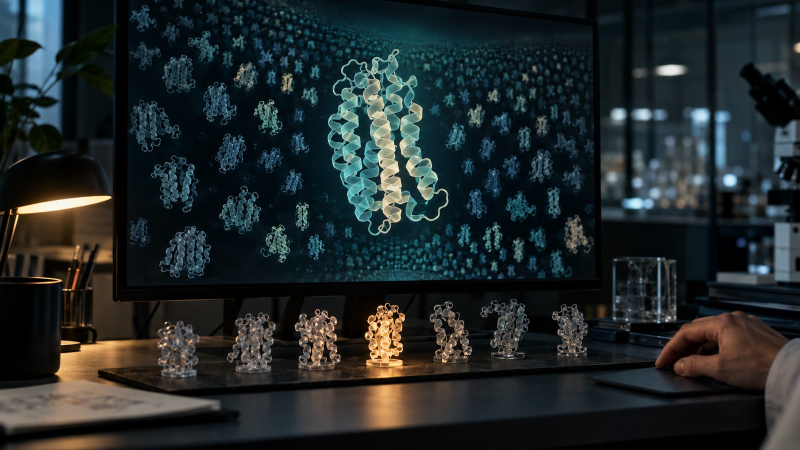
The search began with a molecular silhouette: seven helices bundled through a membrane, the basic architecture of some of biology’s most important receptors.
Researchers at the University of Miami used that shape to search 214,528,851 protein structures predicted by AlphaFold2. They were not asking which proteins had the most similar amino-acid sequences. They were looking for distant relatives whose sequences had changed beyond easy recognition while their three-dimensional forms remained familiar.
One result led them somewhere unexpected.
The protein, TM184C, did not behave like the conventional cell-surface receptor its shape suggested. It appeared mainly inside cells, embedded in mobile vesicles. Under a microscope, those vesicles raced along microtubules, gathered in thin projections and moved between connected cells.
In experiments reported on September 9 in Nature, disrupting TM184C left cultured cells rounded, fragile and short on projections. Adding more of it produced elaborate connections extending tens or even hundreds of micrometres. When differently colored cells were grown together, fluorescent TM184C-positive vesicles crossed between them.
It looks as though a protein hiding in a poorly explored corner of the human proteome helps organize both the cellular recycling system and physical routes for sharing cargo.
There are some hefty qualifications. These were primarily experiments in engineered cells growing in dishes, often using overexpressed and fluorescently tagged proteins. Scientists have not shown what the connections do in an intact animal or human tissue. They do not know whether the exchange helps cells, harms them or sometimes does both.
Even so, the result shows what can happen when AI-generated structures are treated not as answers, but as a searchable map of places biology has barely visited.
Searching by shape
Proteins are chains assembled from amino acids. For decades, one of the main ways to infer what an unfamiliar protein does has been to compare its sequence with those of better-understood proteins. A close match can reveal family relationships and suggest a function.
But sequence is not the whole story. Evolution can substantially rewrite a protein’s amino-acid spelling while retaining a similar fold. Shape-based searches can therefore find connections that sequence comparisons miss.
AlphaFold2 made such searches possible on a previously absurd scale. The system predicts a protein’s three-dimensional structure from its sequence, and the public AlphaFold Protein Structure Database contains hundreds of millions of models.
The Miami team downloaded roughly 24 tebibytes of AlphaFold data and compared the predicted structures against rhodopsin, a classic member of the G-protein-coupled receptor family. GPCRs normally sit in cell membranes and respond to signals such as hormones, odors and neurotransmitters. They are also enormously important drug targets.
The first pass returned more than 1.5 million structural matches. After ranking and classifying them, the researchers singled out six poorly characterized human proteins from the TM184 and PRRT families.
They called this category “superdark.” That is the authors’ term, not an established biological classification. Nor was TM184C literally unknown: it already had database entries, expression data and a tentative connection to cell growth from an older thyroid-cancer study. The mystery was what it actually did.
TM184C was especially tempting because it is broadly expressed and evolutionarily old. It is a 438-amino-acid protein predicted to cross a membrane seven times, much like a GPCR.
But resemblance turned out not to mean identity.


A receptor-shaped protein in the wrong place
A typical GPCR receives a signal outside a cell and activates a G protein inside it. TM184C did not show clear evidence of constitutive G-protein coupling in three assay systems. No ligand is known, and its predicted structure has not been confirmed experimentally.
What it did recruit were β-arrestins and GPCR kinases, two kinds of regulatory proteins closely associated with GPCR biology. The kinases phosphorylated clusters of serine and threonine residues in TM184C’s long C-terminal tail. Those clusters form what researchers call arrestin codes: molecular docking instructions that can influence where a membrane protein goes and what it does.
That tail became one of the study’s most important clues.
Instead of concentrating on the outer cell membrane, TM184C appeared on vesicles roughly 0.5 to 5 micrometres across. The vesicles overlapped with markers for late endosomes, lysosomes and autophagosomes, the compartments cells use to sort, break down and recycle material.
Live microscopy showed them moving in both directions along microtubules. These structural filaments act as intracellular tracks for motor proteins. TM184C-positive vesicles repeatedly split, fused and accumulated at the ends of narrow cell projections.
Some projections met and formed bridges. The authors describe them cautiously as resembling tunnelling nanotubes and tumour microtubes, previously studied structures that can connect distant cells and carry material between them. Whether the TM184C-associated connections should be classified as exactly the same structures remains unresolved.
The genetic experiments make it difficult to dismiss the phenomenon as a colorful microscopy accident. Reducing TM184C with CRISPR produced cells with fewer connections, disorganized vesicles and severe changes in shape. Stable overexpression increased the number and length of projections.
The researchers then disrupted the protein’s tail in two ways. One version lacked its final 82 amino acids. Another retained the tail but replaced 11 potential phosphorylation sites in its arrestin-code regions. Neither variant could restore normal bridge formation after the cell’s endogenous TM184C was removed.
Full-length TM184C could.
That does not yet reveal every step of the mechanism, but it links phosphorylation and arrestin-associated regulation to the physical connectivity of the cells.
Watching cargo change hands
To test whether the bridges were carrying anything, the team grew two engineered cell populations together. One produced TM184C tagged green; the other produced it tagged red. If a green vesicle appeared inside a red cell, or vice versa, the researchers could detect the mixing.
Full-length TM184C moved robustly in both directions. Related proteins TM184A and TM184B did not show the same behavior, and the arrestin-code mutant mostly received vesicles without sending its own back. Incoming normal TM184C-positive vesicles also partly corrected the mutant cells’ abnormal vesicle organization.
The team observed TM184C-positive connections and vesicle exchange in several cultured-cell combinations, including fibroblasts, astrocytes, melanoma cells and patient-derived glioblastoma cells. That variety suggests the behavior is not unique to one laboratory cell line, although every example remains outside intact tissue.
The microscopy also showed TM184C-positive vesicles closely tracking mitochondria, including inside cell-to-cell connections. The paper describes the bridges as supporting organelle sharing, and its supplementary videos show structures associated with mitochondrial transfer.
This is where precision matters. The most direct evidence demonstrates transfer and mixing of fluorescent TM184C-positive vesicles. The mitochondrial observations are intriguing, but they do not establish that TM184C delivers useful energy-producing machinery or provides metabolic support. No experiment in the paper shows what the recipient gains or loses.

The cancer possibilities are similarly open. Glioblastoma cells are known to build interconnected networks, and earlier research has shown that mitochondria can move between astrocytes and tumor cells. TM184C could eventually prove relevant to those systems. The new study does not show that blocking it treats cancer, or even that the protein’s bridge-building behavior occurs in a tumor inside a living organism.
A billion-year detour through yeast
The researchers found a second side to TM184C inside the cell’s waste-processing machinery.
Autophagy packages damaged or unwanted cellular material into autophagosomes, which eventually deliver their contents for degradation. When the team reduced TM184C, levels of lipidated LC3B, a standard marker associated with autophagosomal membranes, increased. Additional inhibitor experiments suggested that the rise came mainly from increased autophagosome production rather than simply blocked disposal.
In other words, TM184C appears to act as a brake or constraint on autophagy under the conditions tested.
The evolutionary test was particularly striking. Yeast has a related vacuolar membrane protein called Hfl1. Previous studies had shown that Hfl1 interacts with the yeast autophagy protein Atg8 and helps maintain the vacuole, an organelle that performs many of the jobs handled by lysosomes in animal cells.
Deleting HFL1 causes abnormal membrane-filled vacuoles. The Miami researchers inserted human TM184C into the deficient yeast, and normal vacuolar morphology returned. Human TM184A and TM184B did not produce the same rescue.
That result supports a deeply conserved role in autophagy-related membrane organization. It does not mean that single-celled ancestors were building bridges between neighbors; the conserved function demonstrated in yeast concerns vacuoles and cellular recycling.
What the AI did, and did not do
The study’s public peer-review record reveals how much work happened after the computational discovery. Reviewers challenged early interpretations and asked for better controls, quantification and rescue experiments. An insufficiently supported narrative about mTOR signaling was ultimately removed. The bridge scoring, automated vesicle analysis and C-terminal rescue experiments substantially strengthened the final paper.
There are still limitations. The bridge measurements pooled image regions from two biological replicates. Experiments were not randomized or blinded, and the phosphosite mass-spectrometry screen was performed once. Most importantly, no independent laboratory has yet replicated a paper published only days ago.
Those caveats do not erase the result. They locate it.
AlphaFold did not look at TM184C and announce that it builds cellular bridges. It predicted structures. Researchers wrote a search, noticed a strange candidate, tested it biochemically, watched it move and repeatedly altered it to see what broke.
Senior author Daniel Isom put the relationship neatly in the university’s account of the work: “AI cannot be blindly trusted, but can lead to really big things in the hands of experts and prepared minds.”
The most interesting part of TM184C may eventually be its role in autophagy, tissue repair, cancer or something nobody has considered yet. For now, it has already demonstrated something valuable: among hundreds of millions of predicted protein shapes, a structural resemblance can be the beginning of an experiment, and an old cellular machine can still have an entirely unexpected story to tell.
Sources / further reading
- Lee et al., Nature, TM184C is a GPCR-like regulator of intercellular exchange and autophagy
- Nature peer review file
- AlphaFold Protein Structure Database
- TMEM184C UniProt entry
- TMEM184C Human Protein Atlas entry
- Liu et al., eLife, Hfl1 and Atg8
- He et al., BMC Biology, Hfl1-mediated vacuolar protein turnover
- University of Miami, Found in the Dark
Keep exploring


