A Few Pancreatic Cancer Cells May Hide Their Neighbors Behind a Fibrin-Rich Niche
A Nature study suggests that rare tumor cells can preserve fibrin-rich pockets that retain suppressive macrophages and keep cancer-killing T cells away. The mechanism is compelling, but the treatment evidence is still confined to mice.

In one experiment, researchers built pancreatic tumors in mice from a deliberately lopsided mixture of cancer cells. Ninety-five percent of the cells had been stripped of a particular immune-evasion gene. Just 5% retained it.
That small minority was enough to change the neighborhood.
The mixed tumors accumulated more macrophages and fewer activated, cancer-killing T cells. By the end of the experiment, the original 5% population had expanded to about 17% of the tumor. More importantly, regions containing even a relatively small share of those cells had already become less hospitable to T cells.
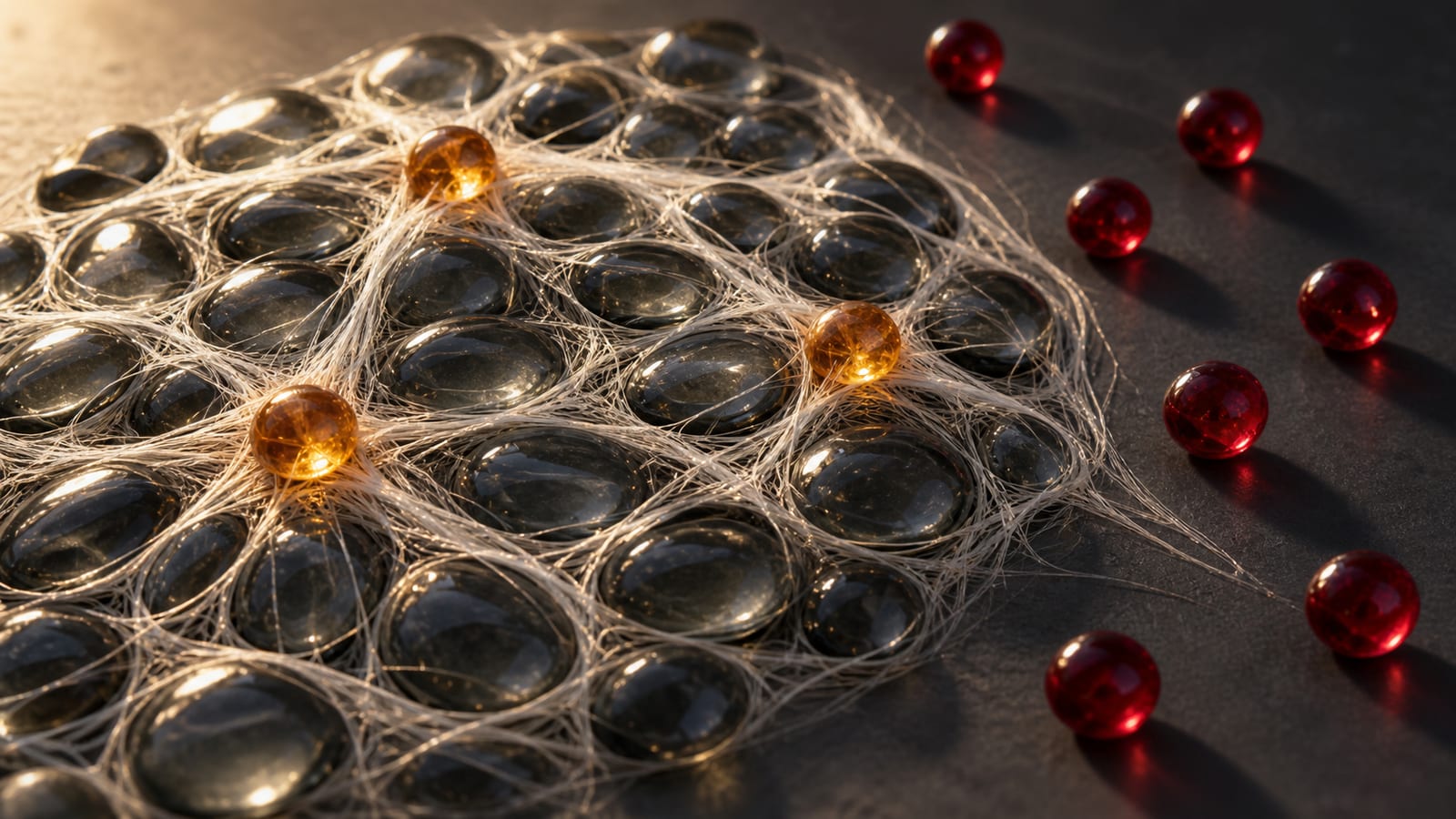
The cells were not merely protecting themselves. They appeared to be building shelter for their neighbors.
A study published in Nature identifies the construction material as fibrin, the mesh-forming protein best known for helping blood clot. Pancreatic cancer cells seem to repurpose this ordinary wound-healing machinery to create localized, immune-suppressive niches.
This is not a conventional blood clot wrapped around a tumor, nor is it simply a wall that immune cells cannot squeeze through. The study suggests something more interesting: fibrin can serve as an active scaffold that attracts, holds and influences macrophages. Those macrophages, in turn, help keep cytotoxic T cells away.
The result is less like a fortress surrounding the entire tumor and more like a collection of hostile little neighborhoods, some organized by a surprisingly small number of malignant cells.
Finding the cells that control the neighborhood
Pancreatic ductal adenocarcinoma, or PDAC, is notorious for the dense mixture of proteins, fibroblasts and immune cells surrounding its cancer cells. This tumor microenvironment is one reason the disease is so difficult to treat.
The five-year relative survival rate for pancreatic cancer across all stages in the United States is about 13%, based on people diagnosed from 2015 through 2021. Immune-checkpoint drugs have transformed treatment for some cancers, but the National Cancer Institute says that a single immunotherapy drug has not been effective for most pancreatic cancer patients. Tumors with microsatellite instability, roughly 1% to 3% of pancreatic cancers, are an important exception.
One possible reason for that broader resistance is simple geography. A checkpoint drug may reactivate T cells, but those cells still need to enter the tumor, remain functional and reach their targets.
The Mount Sinai team studied that geography using Perturb-map, a system that combines pooled CRISPR gene editing, protein barcodes and multiplexed imaging. Each altered population of cancer cells could be identified inside a tumor along with the immune cells in its immediate surroundings.
The initial screen targeted 34 selected genes that encode extracellular or cell-surface factors. Researchers examined more than 4.5 million cancer and immune cells across 20 mice, collecting tumors 7, 14 and 21 days after cancer cells were implanted into the pancreas.


Two genes stood out: SERPINE1 and SERPINB2. They encode proteins called plasminogen activator inhibitor 1 and 2, PAI1 and PAI2.
When either gene was deleted from the cancer cells, the cells still grew normally in a dish. Inside immunocompetent mice, however, tumors formed from the edited cells had less than half the mean burden of control tumors. Their neighborhoods contained fewer macrophages and more CD8 T cells.
That distinction matters. The serpins did not appear to act mainly as an internal accelerator for cancer-cell division. Their advantage emerged from interactions with the living immune environment.
A mesh that does more than block
Fibrin is produced during clot formation and normally cleared through fibrinolysis. In that cleanup system, enzymes called tPA and uPA help convert plasminogen into plasmin, which breaks down fibrin.
PAI1 and PAI2 inhibit those activators. Less plasmin is generated, fibrin breakdown slows and fibrin-rich material can persist.
The researchers found dense fibrin-related deposits in their mouse pancreatic tumors. Removing either serpin reduced those deposits as well as collagen accumulation. Because the antibody used in the tissue experiments can detect fibrin and its precursor, fibrinogen, the paper carefully describes the staining as fibrin(ogen). The wider experimental results support a fibrin mechanism, but that measurement caveat is worth preserving.
Macrophages clustered around fibrin-rich regions and near cancer cells producing PAI1 or PAI2. These immune cells carry an integrin called αMβ2, also known as CD11b-CD18, that can bind fibrin.
The scaffold seemed to affect the macrophages as well as hold them in place. Mouse macrophages cultured on fibrin in pancreatic-cancer-conditioned medium increased production of ARG1 and PD-L1, both associated in this setting with immune-suppressive behavior. Blocking CD18 in tumor-bearing mice reduced suppressive macrophages, increased activated CD8 T cells and slowed tumor growth.
In other words, the fibrin-rich material did not behave merely like inert packing foam. It helped organize and instruct the immune cells occupying the space.
Removing SERPINE1 or SERPINB2 produced substantial immune changes. The knockout tumors contained more than twice as many CD8 T cells as controls. Immunostaining found more than a fourfold increase in CD8 T cells carrying granzyme B, a molecule used to kill target cells. The researchers also measured fewer terminally exhausted T cells.
Those changes made anti-PD-1 treatment more effective in the mouse model. Anti-PD-1 alone had little effect on control tumors. When combined with deletion of either serpin gene, median survival increased from 24 to 47 days.
The team also tested PAI-039, an experimental small-molecule inhibitor of PAI1. Combining it with anti-PD-1 significantly extended survival in two mouse pancreatic cancer models. Blocking CD18 produced a similar improvement in anti-PD-1 activity.
These are encouraging animal results, not evidence that a new treatment is ready for patients. No pancreatic cancer patient received PAI-039 or any other therapy as part of the study.

Do the same neighborhoods exist in people?
The researchers approached that question several ways.
They stained 30 archived human pancreatic tumor specimens and found that fibrin-associated macrophages were located preferentially near malignant cells positive for PAI1 or PAI2. Analyses of three previously published spatial-transcriptomic cohorts also found immune-suppressive, macrophage-rich neighborhoods around serpin-positive malignant regions.
An integrated analysis of five single-cell datasets included 71 patients with enough malignant cells sampled for inclusion. It showed that SERPINE1 and SERPINB2 were usually expressed by discrete minorities of cancer cells rather than uniformly throughout each tumor.
The human data therefore resemble the architecture seen in mice. They do not prove that the pathway causes immune resistance in patients, and they cannot tell us whether blocking it would improve survival.
The finding also joins another recent line of evidence. An independent Science Advances study reported in April 2026 that PAI1 produced by cancer-associated fibroblasts, not just malignant cells, could promote immune-suppressive macrophage states and restrict CD8 T-cell responses in pancreatic cancer models.
Together, the studies suggest that PAI1 may be supplied by more than one part of a tumor. The Nature study's distinctive contribution is showing how rare malignant-cell states can organize fibrin-rich immune niches with effects extending beyond the cells that created them.
What the study does not settle
The authors identify several limitations. Their initial screen covered a curated set of extracellular factors rather than every possible regulator of immune exclusion. They inferred altered fibrin remodeling from tissue deposits and immune effects but did not directly measure fibrinolytic activity or separate the contributions of tPA and uPA. Because the genetic changes were confined to tumor cells, the experiments did not directly test serpins produced by fibroblasts or immune cells.
There is also a practical problem hidden inside the therapeutic promise. Fibrin regulation is part of normal clotting, tissue repair and inflammation. Interfering with it could affect bleeding, thrombosis or wound healing. A useful drug would have to dismantle the tumor's niche without recklessly disrupting the same machinery elsewhere in the body.
Still, the study changes the picture of what immune resistance can look like. A tumor does not need every malignant cell to run the same defensive program. A small faction may alter the terrain for everyone nearby.
That may be the most consequential finding: inside a heterogeneous tumor, power is not always proportional to population. Sometimes 5% is enough to redesign the neighborhood.
Sources / further reading
- Nature, A serpin-myeloid axis in pancreatic cancer heterogeneity and immune evasion
- National Cancer Institute, Advances in Pancreatic Cancer Research
- American Cancer Society, Survival Rates for Pancreatic Cancer
- Science Advances, A stromal PAI1-tPA axis orchestrates immunosuppression in pancreatic cancer
- Mount Sinai, Pancreatic Cancer Uses Blood-Clotting System to Evade Immune Attack
Keep exploring


